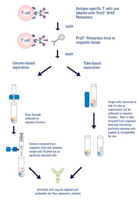
The red classification laser (635 nm) or LED interrogates the internal dyes to identify bead regions. Dyed beads are pushed through a detection chamber in a single file or magnetically immobilized.
#BIORAD MAGNETIC BEADS ANTIBODY REACTIVITY SOFTWARE#
In all readers, high-speed digital signal processors and software record the fluorescent signals simultaneously for each bead, translating the signals into data for each bead-based assay (Figure 2).įig 2. Classification and reporter excitation is accomplished with the use of LEDs rather than lasers. With the LED/image-based Bio-Plex MAGPIX reader, the beads are drawn into a chamber and magnetically immobilized. The green reporter laser excites the reporter molecule associated with the bead, which allows quantitation of the captured analyte. The red classification laser excites the dyes in each bead, identifying its spectral address. For the flow cytometry–based systems, precision fluidics align the beads in single file through a flow cell where two lasers excite the beads individually.

Each bead region is conjugated to a specific target analyte (a) followed by binding with a biotinylated detection antibody (b) and a reporter dye, streptavidin-conjugated phycoerythrin (c).ĭuring data acquisition, the contents of each microplate well are drawn into the array reader, depending on the type of reader - either the flow cytometry/laser excitation–based Bio-Plex 200 and Bio-Plex 3D systems or the light emitting diode (LED)/image-based analysis employed in the Bio-Plex ® MAGPIX™ multiplex reader. Different concentrations of red and infrared dyes are used to generate up to 100 distinct bead regions.

Beads are colored internally with two different fluorescent dyes (red and infrared). A dual detection flow cytometer is used to sort out the different assays by bead colors in one channel and determine the analyte concentration by measuring the reporter dye fluorescence in another channel (Figure 1).įig 1. The use of different colored beads enables the simultaneous multiplex detection of many other analytes in the same sample. The technology enables multiplex immunoassays in which one antibody to a specific analyte is attached to a set of beads with the same color, and the second antibody to the analyte is attached to a fluorescent reporter dye label. The reagents may include antigens, antibodies, oligonucleotides, enzyme substrates, or receptors. These beads can be further conjugated with a reagent specific to a particular bioassay. This technique involves 100 distinctly colored bead sets created by the use of two fluorescent dyes at distinct ratios. The Bio-Plex ® multiplex immunoassay system utilizes xMAP technology licensed from Luminex to permit the multiplexing of up to 100 different assays within a single sample. The output of the Human Genome Project, for example, provides the ability to simultaneously monitor the roles of multiple genes during investigations of complex biological systems. The growth of proteomics and genomic analysis is driving the need to discover and monitor large numbers of biomarkers indicative of human disease states. The impact of immunoassays on life science research and clinical diagnostics has been enormous, with almost 10,000 studies published per year that include the terms “enzyme immunoassay” and “enzyme-linked immunoassay” (Lequin 2005).

Today, key advantages of ELISA are its ease of use, flexibility, and low cost. While early versions did not rival the sensitivity of the RIA, the development of highly specific monoclonal antibodies and chemiluminescence detection resulted in ELISA assays with sensitivity that exceeds that of radiolabels. ELISA uses an enzymatic reaction as the basis of detection, rather than a radioactive signal. The search for a suitable alternative to the RIA led to the development of ELISA in the early 1970s ( Engvall and Perlmann 1971, Van Weeman and Schuurs 1971). The radioimmunoassay (RIA) would remain the standard for the detection of bioanalytes for more than ten years because of its extraordinary sensitivity, despite the health risks and disposal issues posed by the use of radioisotopes. These initial assays used radiolabels for detection. The first immunoassay was developed by Yalow and Berson (1960), who received the Nobel Prize for their efforts to measure insulin levels.


 0 kommentar(er)
